Setting up the meso-veins protocol at 7T.
Implementing the 0.35 mm isotropic whole brain multi-shot multi-echo 3D-EPI protocol (a.k.a. the mesoveins protocol) at a new scanner (Siemens 7T) requires combination of a custom pulse sequence and parameter replication. While we have mentioned aspects of this in our paper (Gulban et al., 2026), here is the detailed workflow.
1. Access the Pulse Sequence
The foundation is the custom multishot 3D-EPI sequence compiled by Rüdiger Stirnberg (DZNE). You can access this sequence via the Siemens C2P exchange platform: https://webclient.eu.api.teamplay.siemens-healthineers.com/c2p and search for these terms to find the package:
3D EPI,3D Echo Planar Imaging,DZNE Bonn,dzne_ep3d
2. Protocol Replication
Once the sequence is available on your scanner, you can replicate our specific protocol using the parameter PDF archived on Zenodo: https://doi.org/10.5281/zenodo.14145584. Download ME3DEPI_pt35_whole_brain_3echos_7min_UPDATE-v3p1.pdf for the latest updated example protocol.
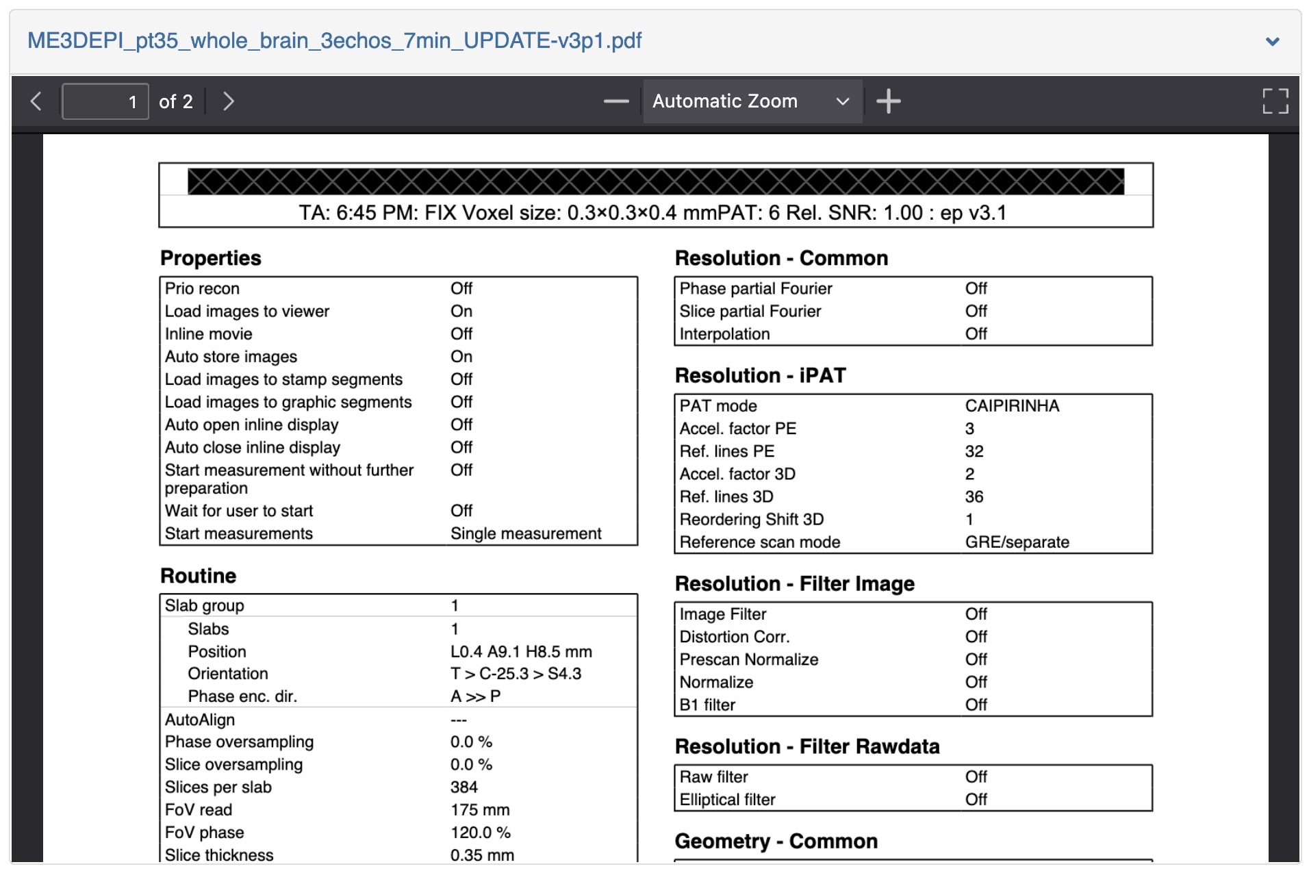
Note that we have our previous protocol that was used to acquire the publised data during 2024-2025 years (named ME3DEPI_pt35_whole_brain_3echos_7min.pdf), however Rüdiger developed the sequence further since then. Therefore, currently, we advise you to copy the parameters from ME3DEPI_pt35_whole_brain_3echos_7min_UPDATE-v3p1.pdf.
If you get stuck at while replicating the scanning parametres, try following the steps below:
- Temporarily reduce contrasts to 1.
- Temporarily increase TE1 to about 15ms.
- Reduce base resolution to 500.
- Temporarily reduce readout bandwidth.
- Set FOV read: 175mm (along L>R).
- If that is not possible immediately, keep on reducing the readout bandwidth until it works.
- Set FOV phase: 120% (that gives matrix size 600 that is 210mm along A>P).
- Set Slices per slab to 370, with 0.35 mm slice thickness.
- Increase readout bandwidth and undo all other temporary changes (Minimize TE1, increase the number of TEs to
- minimize TR).
- If you get PNS error, add 0.5 ms to TE1 until you don't get the PNS error.
3. Test on a phantom
While we aim for high-fidelity replication, site-specific hardware may result in slightly different parameter offsets. Do not be concerned if settings are not a 100% match. However, it is strongly recommended to test the newly configured protocol on a phantom first to ensure the sequence completes and reconstructs successfully. The meso-vein protocol generates a significant voxel load. You must monitor the reconstruction duration relative to the acquisition time:
- Acceptable: 7-minute scan followed by 6 minutes of reconstruction.
- Critical: 7-minute scan followed by 15+ minutes of reconstruction.
If reconstruction time significantly exceeds scan time, your reconstruction PC may become "clogged", potentially leading to system failure during back-to-back scans. If you observe persistent reconstruction lag, contact your site technician.
4. Test on a Human & Slab Positioning
Following a successful phantom session, you can proceed to in vivo acquisition. Proper slab orientation is critical for managing specific 7 T artifacts. As illustrated below, the imaging slab is tilted to approximate the AC-PC (anterior commissure, posterior commisure) plane. This positioning ensures full cerebellar coverage while strategically shifting skull bone and fat-wrapping artifacts into nasal cavity in between the brain stem and inferior frontal cortex. Also, it is critical to avoid the eyeball as much as possible as they can cause 3D EPI artifacts particularly if the participant is moving their eyes a lot or blinking frequently and strongly.
5. How to instruct the participants for the best mesoveins images
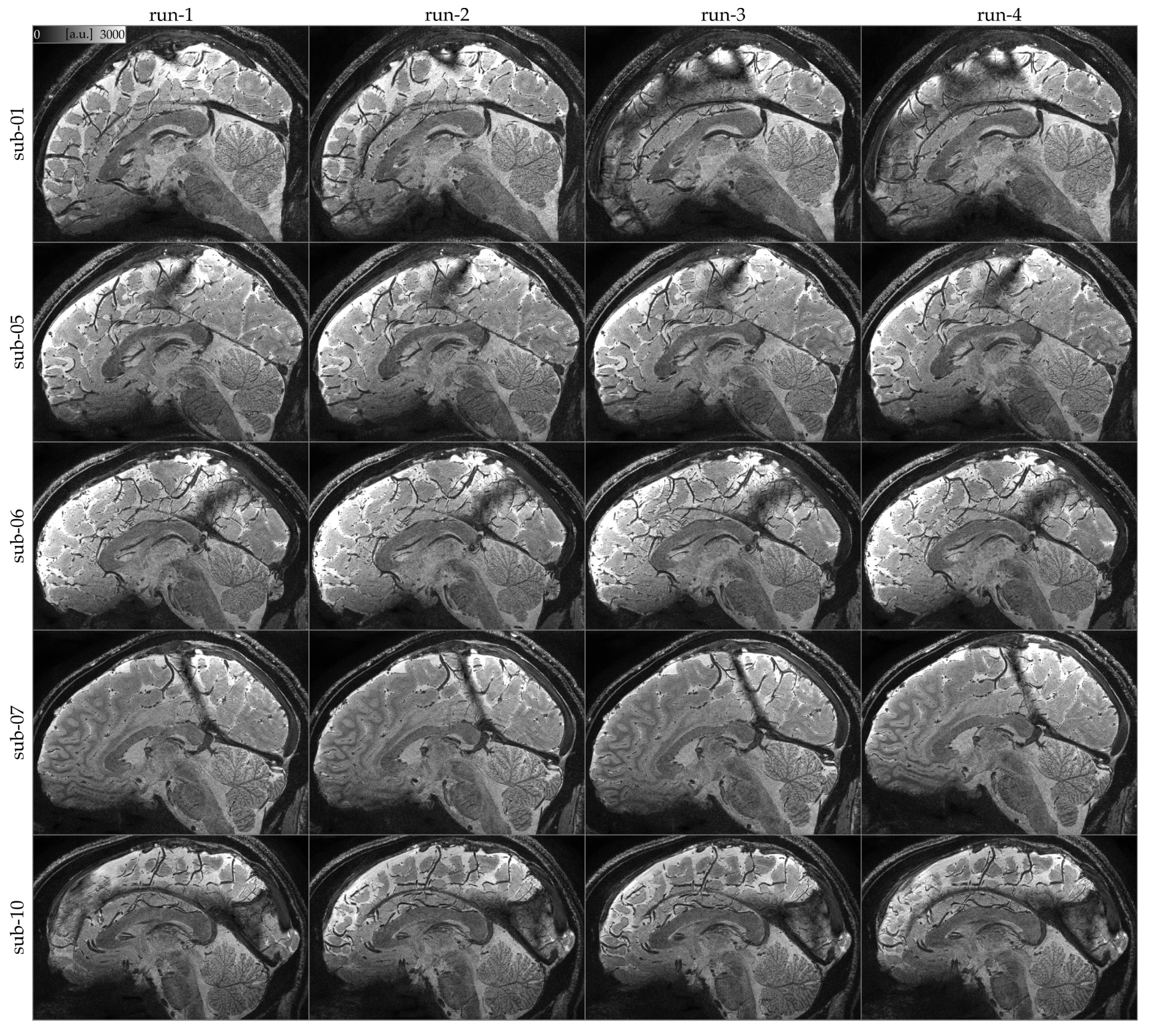
We observed that strict instructions often lead to worse results. During breaks between runs, we explicitly instructed participants: "Please feel free to slightly move your head now if needed.". In our experience, providing these "micro-releases" significantly reduced involuntary bulk head motion during the actual scan compared to the conventional instruction to stay perfectly still for the entire hour. In one case (sub-07 run-04 above), the participant moved for several millimeters in between the runs, but managed to stay still within the acqusition. We could easily recover this data by using rigid body motion correction in post processing.
Next post will be about quality controlling the meso-veins images, particularly in the context of head motion artifacts. Stay tuned.
Acknowledgements
These tips are distilled from extensive technical exchanges with Rüdiger Stirnberg, alongside invaluable hours of collaborative scanning with Dimo Ivanov. I am particularly indebted to our coffee break conversations with Renzo Huber, his insights into acceleration parameters served as the catalyst for this work. My thanks also to Alessandra Pizzuti for her critical support in navigating the parameter conflicts when setting up the protocol. I am deeply grateful for their generosity in sharing their expertise and for the collective effort required to refine this meso-vein protocol.